Like a Circus: The Public Consumption of Sex Differences
Claim
Source
Boys’ brains develop from the back of the head to the front, from the ‘doing’ part of the brain to the ‘thinking’ part, whereas girls’ brains develop from the front of the brain to the rear. This means that boys are able to act before they are able to think
Hodgins (2011)
The resting female brain is more active than the male brain, which often goes into a pause state after tasks. To break the pause, boys must use loud voices, run, or jump
Boys don’t remember what you have told them. Each time an incident happens, it’s as if it has never happened before
Hodgins (2007)
Girls’ brains experience 15 % more blood flow than boys’
Girls tend to use the more advanced parts of their brains, whereas boys use the more primitive parts
McBride (2008)
Girls develop language 6 years earlier than do boys
McBride (2008)
The corpus callosum is 20 % larger in girls than boys. This means that boys have trouble talking about their emotions, since emotion and language are located on opposite sides of the brain
Boys have half as much neural tissue devoted to verbal-emotive functioning
McBride (2008)
Boys have less oxytocin than girls, which makes them uncomfortable with eye contact. They should be seated side-by-side, to avoid such
Boys have less serotonin than girls, which makes them more fidgety and impulsive
Girls can hear better than boys
Girls can see better in dim light
Chadwell (2010)
Boys’ visual systems are wired to detect moving objects
Girls’ visual systems are wired to respond best to the colors red or pink
Chadwell (2010)
Boys are most comfortable at a temperature of 69 °F whereas girls work best at 75 °F
Sax (2006)
Girls are able to see the details of a situation because the detailed area of the brain, called the crockus, is four times larger in girls than boys
Hodgins (2007)
The effect of such presentations on educational policy in the US was stunning. In 2012, the American Civil Liberties Union reported that of the single-sex education programs they investigated, nearly all cited pseudoscientific material from the popular press, not peer-reviewed literature (see Table 1), as justification for separating the sexes (American Civil Liberties Union 2012). In order to accommodate what they believed were scientifically proven sex differences , schools used different colors to decorate the classrooms, set thermostats at different temperatures, and arranged seating with girls face to face to promote social interactions and boys side by side to avoid eye contact. In an all-boys classroom in Idaho, teachers used microphones to adjust their voices to a level they were told is best for boys (Hollingsworth and Bonner 2012). Teachers at a school in West Virginia were told that girls need low light levels; the lighting was so low in a girls’ classroom that a visually impaired student could not see well enough to function (Khadaroo 2012). In some cases, parents sued to end mandatory single-sex instruction (e.g., Doe 2012) but were not always successful (e.g., A.N.A. 2011).
The use of pseudoscience to justify these new practices triggered a strong response from scientists and gender studies scholars. Several critical books and articles were published between 2009 and 2011 (Eliot 2009, 2011; Fine 2008, 2010; Halpern et al. 2011; Jordan-Young 2010; Rivers and Barnett 2011). Work remains to be done, however, as proponents of single-sex classrooms continue to perpetuate myths and stereotypes, and school administrators continue to listen. Those myths and assertions (see Table 1) have been thoroughly debunked elsewhere; my goal in this article is instead to consider the ways in which we have failed to adequately communicate the nature of sex differences to the public and to suggest ways in which we might help teachers and parents better evaluate them. First, we need to recognize that after our findings are published in scholarly journals, they are filtered and sensationalized by a series of non-expert translators, such as the popular media and teacher educators (Hardiman et al. 2012). Sex differences are packaged and sold to schools as evidence that boys and girls fall into dichotomous categories with non-overlapping distributions. Certainly, small differences do inform our understanding of the factors that contribute to learning and their value should not be discounted—yet, as scientists, we are obligated to respond to misrepresentation of our findings to promote a social agenda, and to establish a more effective dialog with policymakers. In addition, we need to address our own propensity to draw illogical inferences about the meaning of sex differences, particularly from neuroimaging data. Ultimately, because sex differences are so easily misunderstood and misinformation potentially harmful, we need to hold others and ourselves to a high standard when reporting them.
2 What is a Sex Difference?
Everyone understands intuitively that the sexes are different, because our sex organs are obviously different. With few exceptions, a child is categorized as one sex or the other from the moment he or she is born. Because of the widely recognized differences in genitalia, it is easy to believe that other differences between the sexes could be equally large. MRI technology affords unprecedented views inside areas that historically have been obscured from view. If educational consultants argue that newly discovered sex differences in the brain and behavior are large and meaningful enough to warrant different classrooms for girls and boys, teachers often listen.
Actual sex differences in behavior and the brain, particularly in children, should certainly not compel educators to implement dramatic new policies. For example, consider sex differences in impulsivity or activity, the effect size (Cohen’s d) of which is typically about 0.2 (Hyde 2014). A hypothetical effect of this magnitude is plotted in Fig. 1a. Such sex differences, which might instead be called ‘sex effects’ to avoid the term ‘difference’ altogether, can be statistically significant, but only when sample sizes far exceed a typical elementary school class. In other words, such an effect would not be detectable within a class, a grade, or possibly even an entire school—an effect of the size depicted in Fig. 1a would require approximately 400 children to detect. Because distributions are almost never plotted in research reports, the degree of overlap between the sexes is usually lost when the finding is communicated to teachers. As a result, effects like the one in Fig. 1a are presented and interpreted as evidence that boys and girls cannot learn optimally in the same classroom. Yet, for every 50 boys above the mean, there are 46 girls also above it. Similarly, for every 50 girls below the mean, there are 46 boys below it. Thus, if it is true that children above and below the mean need different classroom environments, separating them according to their sex would do very little to address that need. Almost as many children would end up in the ‘wrong’ classroom as in the ‘right’ one. Such a strategy, which would benefit only the children at the extremes of the distribution, essentially constitutes teaching to the tails; it does not consider the needs of the majority of the children, for whom sex does not predict ability or behavior.
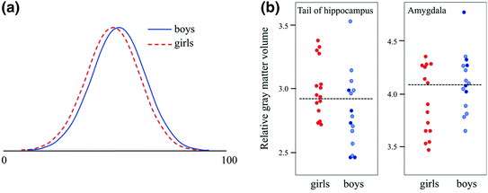
Fig. 1
Sex ‘differences’ in behavior and the brain typically show large overlap. (a) Normal distributions showing a typical sex difference in a behavior or personality trait, such as impulsivity or activity (reviewed by Hyde 2014). The number of boys or girls with scores on a hypothetical scale of 0–100 is plotted. For every 50 boys above the mean, there are also 46 girls above the mean; the sexes overlap by more than 80 %. This difference (effect size d = 0.2) is actually much larger than those typically reported for traits such as verbal or mathematic ability. Even for traits with larger sex differences , for example, interest in things vs. people (effect size = 0.93), the overlap is close to 50 %. (b) The size of the hippocampus and amygdala varies according to sex in children ages 8–15 (data from Neufang et al. 2009). If this sample of 30 children were split according to the median size of either structure (dashed lines), a large proportion of the children would be in the ‘wrong’ group for their sex. The ‘small hippocampus’ group would consist of 9 boys and 6 girls; the ‘small amygdala’ group would consist of 7 boys and 8 girls. Notably, each of the ‘girl-like’ groups would contain one or two boys with testosterone levels typical of mid-puberty
A more concrete example of overlapping distributions appears in Fig. 1b, which depicts a known sex effect on the sizes of two brain regions. According to Neufang et al. (2009), the hippocampus is larger in girls and the amygdala larger in boys. Educational consultants have used such findings to argue for large sex differences in information processing and emotive functioning (Gurian and Stevens 2004, 2005; Sax 2005). A close inspection of the actual data reveals large overlap; if we were to use the median hippocampus or amygdala size to divide the students into groups, the number of boys and girls would be approximately equal in each. The authors of the study found that the surge of testosterone in pubescent boys may explain the larger amygdala; importantly, our class with ‘girl-like’ amyg-dalae would even contain two older boys that had begun puberty. Thus, although these brain structures are different in that an effect can be detected, sex is a rather poor predictor of their size. Certainly, if a small hippocampus and large amygdala warrant a certain educational approach, dividing students by sex would not be a good strategy by which to implement that approach.
Neuroanatomical and psychological data almost never fall into distinct clusters corresponding to sex (e.g., see Carothers and Reis 2013). For this reason, looking to sex as a source of ‘difference’ in the brain has been criticized (Jordan-Young and Rumiati 2012). But because most people regard sex as a category rather than as a continuous variable, even the smallest effects are an easy sell. Single-sex education programs offer a convenient solution to a vexing, urgent problem. Just as Men are from Mars, Women are from Venus (Gray 1992) promised to improve relationships by showing us how to embrace difference, single-sex classrooms promise to save our failing school system. We will need better ways to convey to the public that boys and girls are in fact the same—not from Mars and Venus or even, as some have phrased it, from North and South Dakota (see Eliot 2009). Looking at Fig. 1, I would argue that although a couple of boys may hail from Hoboken and one or two girls from Hackensack, the rest of the children are all from New York City.
3 When a Difference is not a Difference at All
Many sex differences reported in the media and presented to school administrators have uninteresting explanations or no support at all. Here, I will discuss just one example: the amounts of gray and white matter in the brain. Most of the relevant studies suggest that the average amount of gray matter in women is slightly higher than in men (reviewed by Cosgrove et al. 2007). Gray and white matter volumes are closely tied to overall brain volume, which is about 10 % larger on average in men than women. When gray matter volumes are corrected for overall brain size, the sex effect is substantially lessened (Leonard et al. 2008) or eliminated (Blatter et al. 1995; Courchesne et al. 2000). As an example, data from Lenroot et al. (2007) are shown in Fig. 2a and then redrawn corrected for brain volume in Fig. 2b. The relative amounts of both gray and white matter in children ages 7–19 appear to be exactly the same in boys and girls.
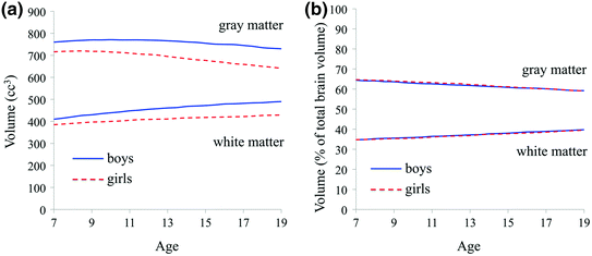
Fig. 2
Some sex differences in the brain may be explained by overall brain size, which is larger in boys. (a) Data from Lenroot et al. (2007) suggest that levels of both gray and white matter are higher in boys. (b) When the average values are divided by the average values given for overall brain size, the sex difference appears to be eliminated
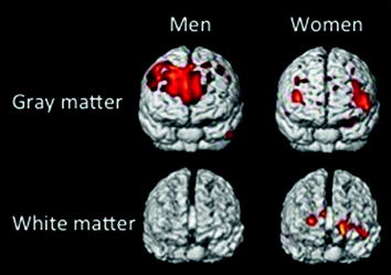
Despite the large literature showing that the sexes have similar amounts of gray and white matter, professional development materials for teachers often paint a starkly different picture. They routinely assert that boys have 6.5 times as much gray matter as girls, and girls have a whopping ten times as much white matter as boys (Box 1). A difference that large would be obvious using techniques available before the dawn of recorded history (e.g., looking at the brain of a deceased person), and would be just as salient as sex differences in reproductive organs. As of this writing, a Google search on ‘women have 10 times more white matter’ produces more than 6,000 hits. The CBS Early Show covered this difference as if it were breaking news (CBS News 2010) but provided no source. With some effort, I traced the myth to media coverage of a paper by Haier et al. (2005). As I expected, these authors did not report sex differences in the amounts of gray or white matter. Rather, they used structural MRI to identify areas of gray and white matter in each participant’s brain and then searched for correlations between the sizes of those areas and scores on an intelligence test. The measures that were 6.5 times larger in men and 10 times larger in women were not the total gray or white matter volumes, but rather the volumes that predicted the score on the test—without regard to whether those correlations were statistically significant. The scores were significantly related to the size of only a few areas of gray matter in men, and one in women. In a press release, the authors commented that human evolution has created two different types of brains designed for equally intelligent behavior (Today@UCI 2005). The media then proceeded to run amok, giving rise to perhaps the most nonsensical neuroscience myth since the one about humans using only 10 % of their brains. If Haier et al. have attempted to address the confusion, their attempts have been swamped by the sheer volume of misrepresentations.
Box 1 An urban legend is born. A 2005 paper by Haier et al. was so grossly misinterpreted by the media that it gave rise to a now-pervasive urban legend: Men have 6.5 times as much gray matter as women, and women have 10 times as much white matter as men. The 2005 paper, which described a structural MRI study relating intelligence to certain voxels of gray and white matter in men and women, contained a version of the figure above. The figure has been reproduced hundreds of times on the internet, sometimes with an accurate caption but more often with a caption such as, “Activity in men and women while taking an IQ test” (no imaging was actually done during the test) or “Men have 6.5 times as much gray matter and women have 10X as much white matter”. Figure from Andrew-Sfeir (2012).
4 Leaps of Logic and the Allure of the Brain Scan
Proponents of brain-based single-sex education have argued that modern imaging techniques have revealed large differences between the brains of boys and girls (Chadwell 2010; Gurian and Stevens 2005). They present images of the BOLD response or white/gray matter distribution, chosen to illustrate differences that may or may not have ever been reported in peer-reviewed literature. Such images are quite powerful; one teacher wrote, ‘I was trained in the idea that each student is an individual. But when I saw the PET scans of boys’ and girls’ brains, I saw how differently those brains are set up to learn.’ (Gurian and Stevens 2004; italics added). Note that in addition to believing that the images represented a typical boy and girl, the teacher was convinced that they showed something about learning styles. Although that leap of logic is a large one, it is common. Such ‘reverse inferences ’ (Poldrack 2006) rest on the fallacy that if neuroanatomical differences exist, they must explain behavioral differences. The larger hippocampus of girls, according to materials distributed to teachers, endows them with better memory, social skills, and language skills (Gurian and Stevens 2004; McBride 2008). Similarly, the larger amygdala of boys supposedly makes them more aggressive, reduces their attention span, and increases the amount of space they require in the classroom (Gurian and Stevens 2005; Multiplying Connections 2012). The scientific basis for these claims is unclear, but to the non-expert, they apparently seem plausible.
Perhaps the most pervasive of illogical reverse inferences is the attribution of cognitive abilities to the relative volumes of white and gray matter, which is presumed to be related to the degree of interconnectedness among brain regions. The evidence for sex effects on both white matter volume and connectivity has been reviewed elsewhere (e.g., Bishop and Wahlsten 1997; Bruner et al. 2012) and is tangential to my point here: How and whether these factors affect abilities is unknown. Sex differences in connectivity and white matter volume have nonetheless been cited as evidence of either male or female superiority in spatial orienting, language skills, empathizing, map reading, mathematics, and multitasking (reviewed by Fine 2010). To a non-expert, the absence of a known function may not be particularly relevant because a sex difference implies function. For example, if women are found to have more white matter and men more gray matter, then white matter is said to be responsible for multitasking and gray matter confers mathematical ability (CBS News 2010). Conversely, if men are found to have more white matter and women more gray matter, then white matter is reported to confer mathematical ability, while gray matter is important for multitasking (Chamberlain 2009). Ignored are the findings that mathematical ability does not vary with sex (reviewed by Hyde 2014), and the only two studies on multitasking showed no female advantage (Hambrick et al. 2010; Mäntylä 2013). A sex difference in the brain appears to be enough to convince many people that a difference in ability must exist, despite the maddening circularity of the logic.
Why are such tenuous arguments so convincing? First, they support stereotypes. The combination of reverse inference and social stereotypes is a dangerous one, as was famously illustrated by Gould in The Mismeasure of Man (1981). Gould debunked nineteenth-century research alleging that intellectual superiority of white men could be explained by their larger cranial capacity, compared with women and men of other races. Gould argued that the conclusions of the researchers were shaped by their own expectations. The stereotypes of that century ensured that the research would be accepted, even embraced. Likewise today, brain-based explanations for effects of sex on achievement capitalize on long-held stereotypes, often triggering aha moments for parents and teachers as they become convinced that their own personal beliefs are validated by science (Kaufmann, n.d.). The more dearly held those beliefs, the harder it is to convince the believer that such arguments are flawed. As one New York Times reader commented, ‘[Feminists] just love to pretend there are no hard differences between the brains of men and women,’ which the reader called a ‘brazen denial of what is not only real but thunderingly obvious’ (comment on Schott 2010).
Sadly, the use of reverse inference to perpetuate stereotypes is not limited to amateurs. Neuroscientists themselves are guilty (see Bluhm 2012). In a recent imaging study, Ingalhalikar et al. (2014) described effects of sex on the ‘connectome’ of the brain, arguing that women showed stronger interhemispheric connections, whereas men’s brains were better connected within hemisphere. In the discussion section of the paper, the authors inferred that the male-typical pattern would confer an efficient system for coordinated action, whereas the female-typical interhemispheric connections would better integrate the ‘analytical’ left hemisphere with the ‘spatial and intuitive’ right hemisphere. The degree of overlap between the sexes was not reported; in the institution’s press release, however, the authors described the sex difference as “stark” and “striking” and suggested it might explain why men are better at cycling and women better at socializing and multitasking (Penn Medicine 2013). In an interview, an author remarked, ‘I was surprised that it matched a lot of the stereotypes’ (Sample 2013